
Description
Specifications
Documents
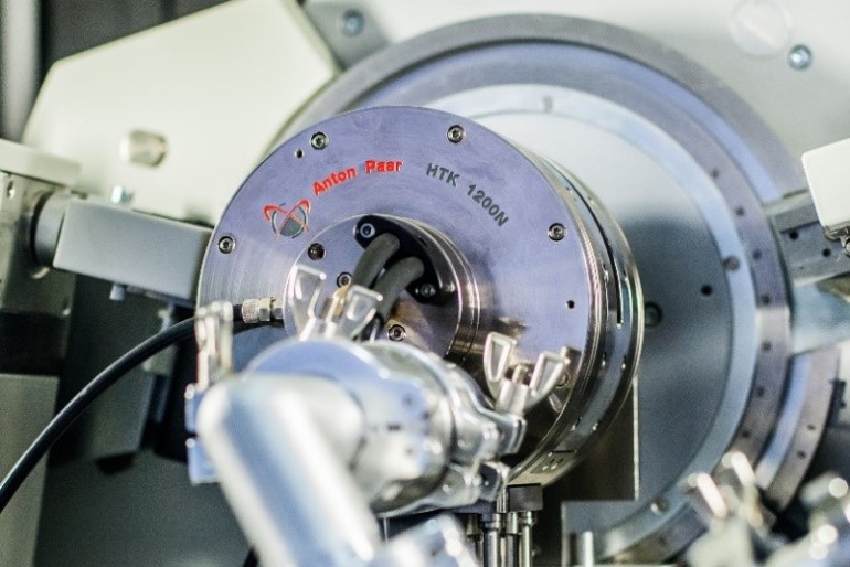
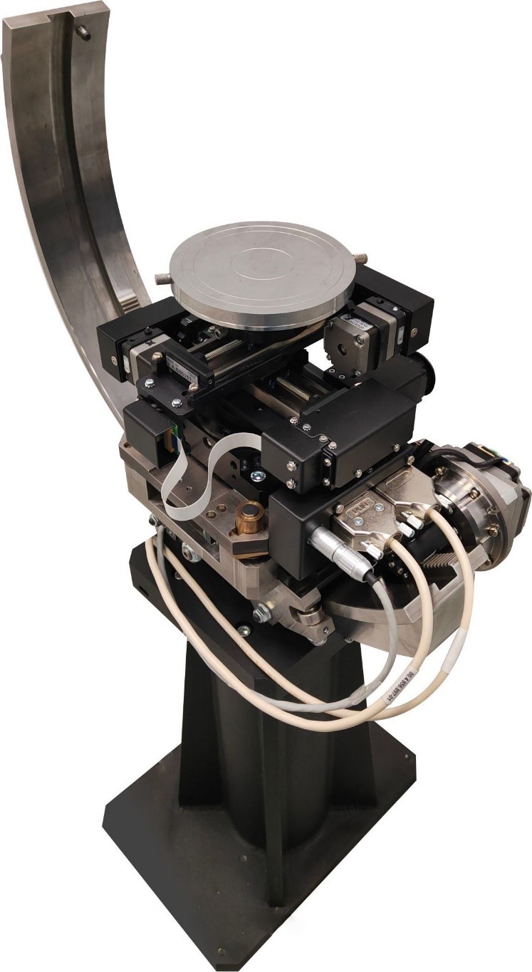
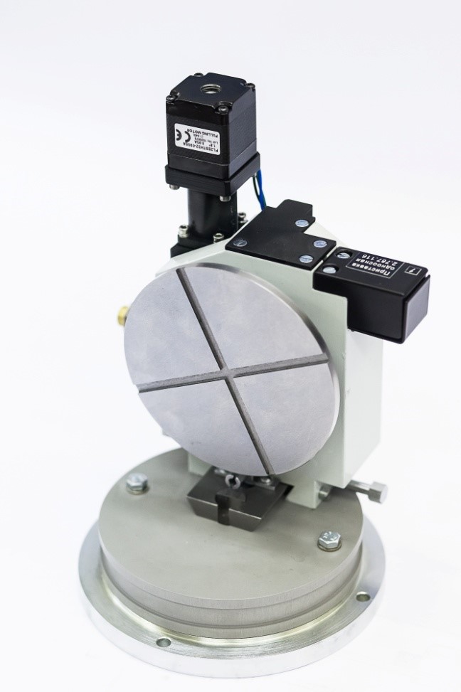
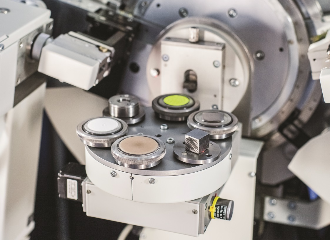
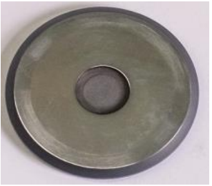
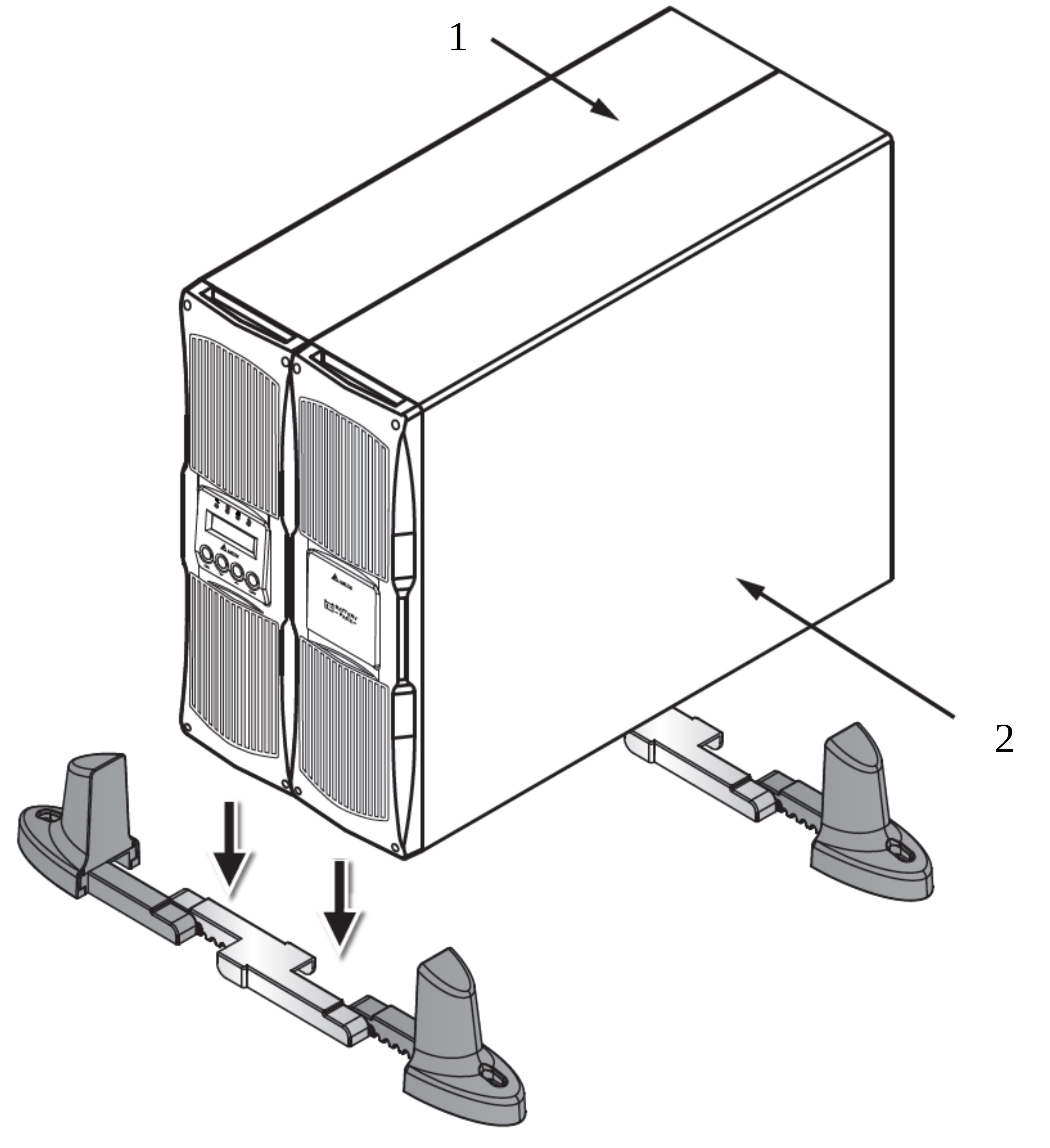
Basic equipment
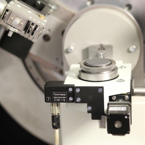
Options
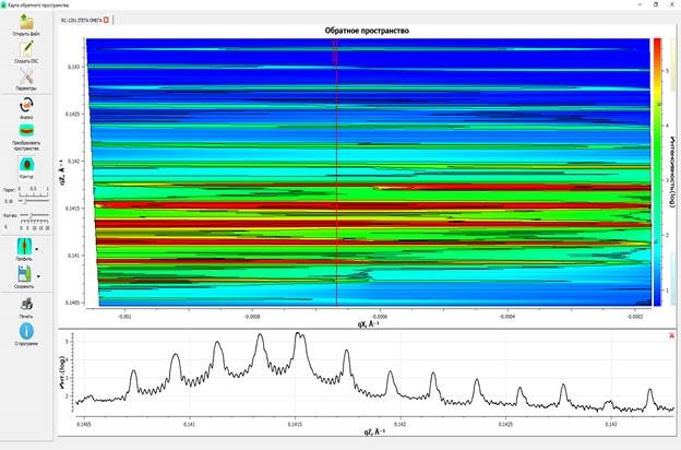
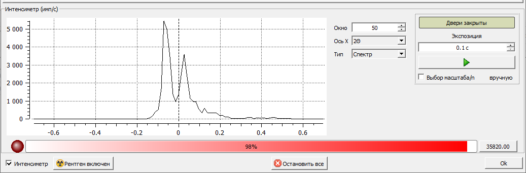
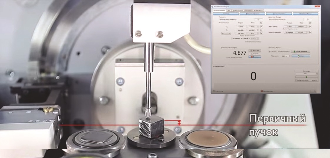
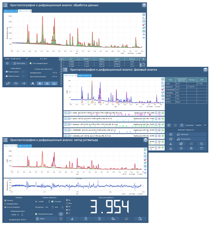
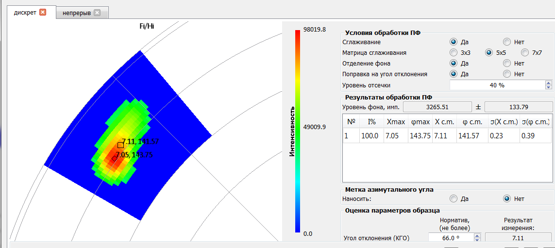
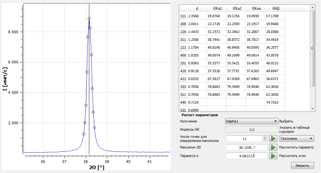
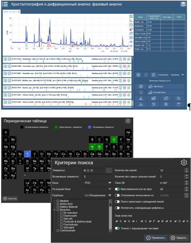
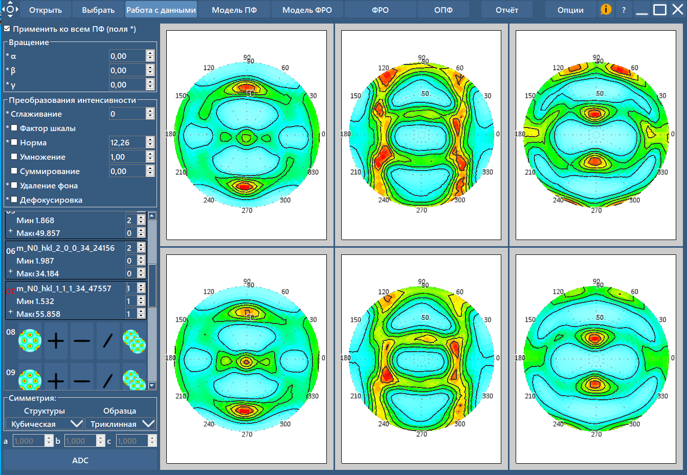
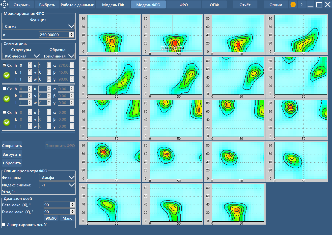
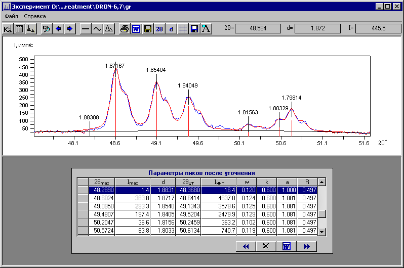
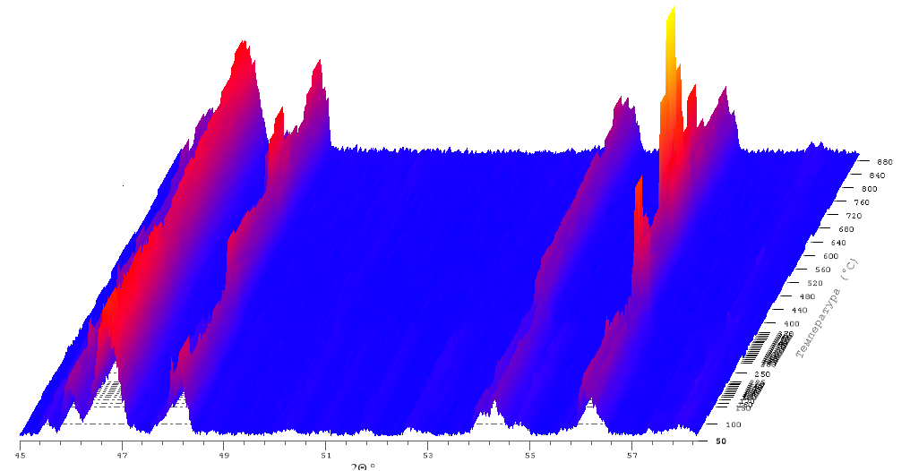
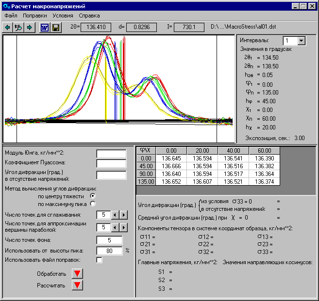
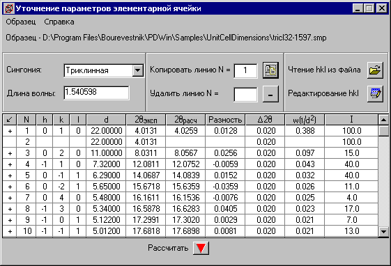
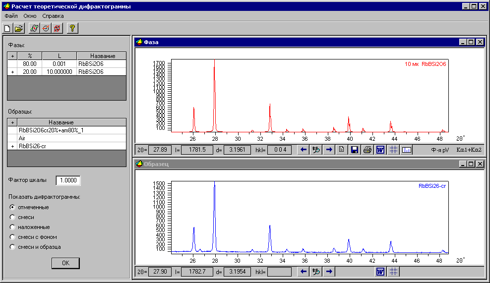
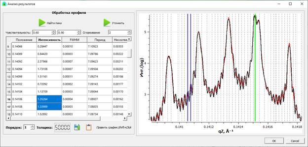
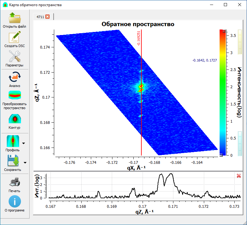
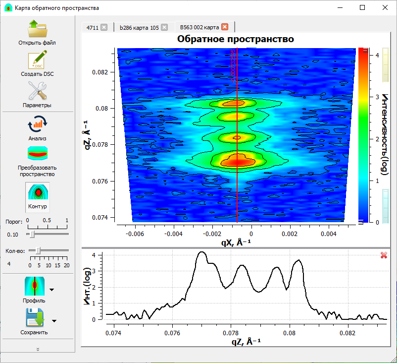
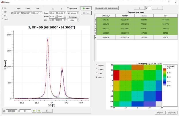
Software
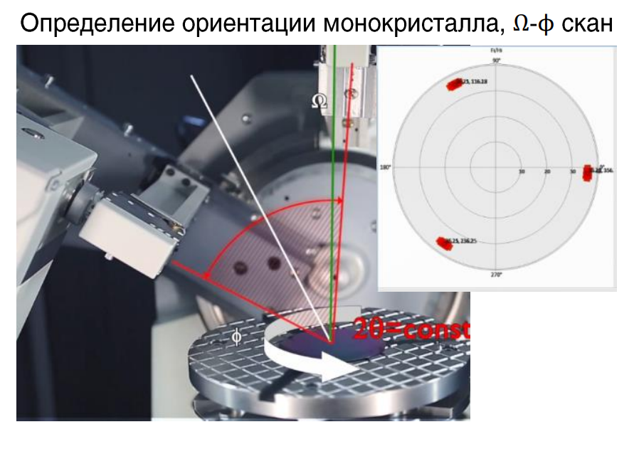
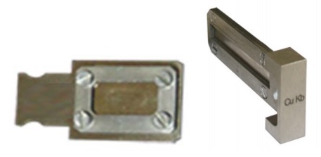
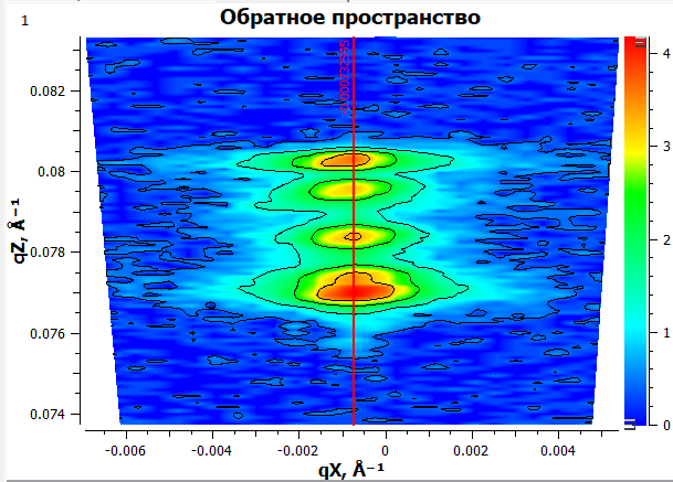
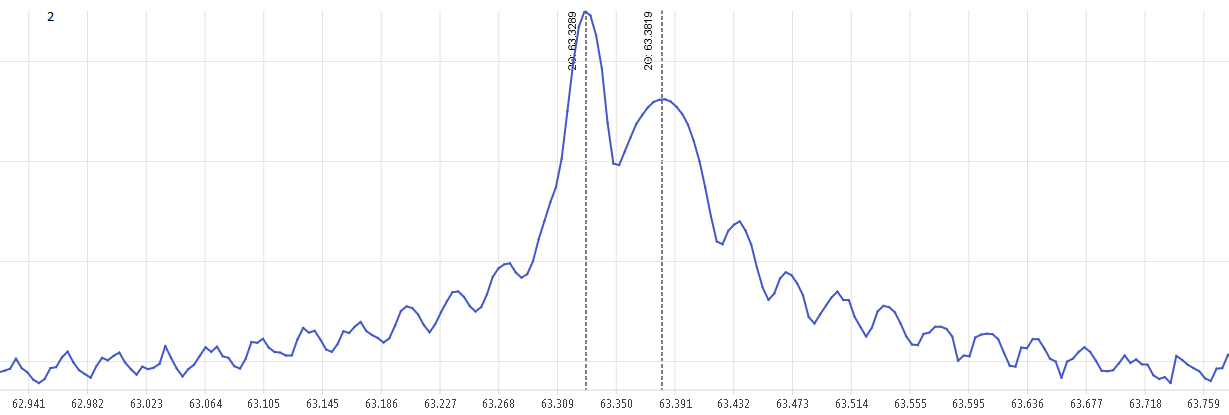
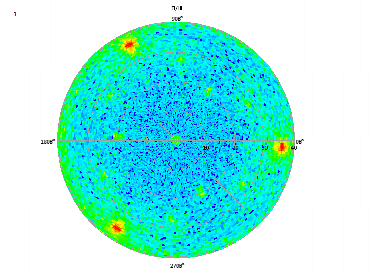
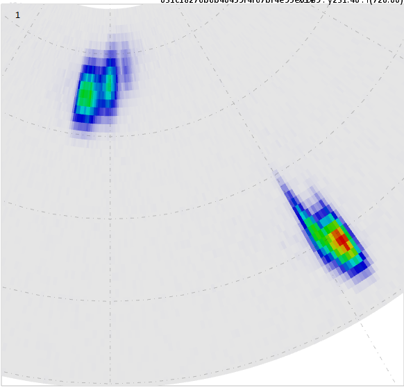
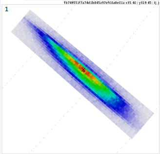
X-ray optical patterns
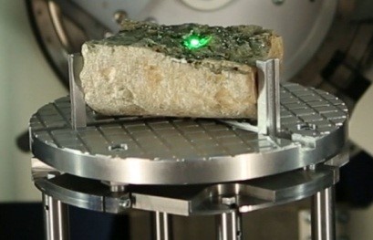
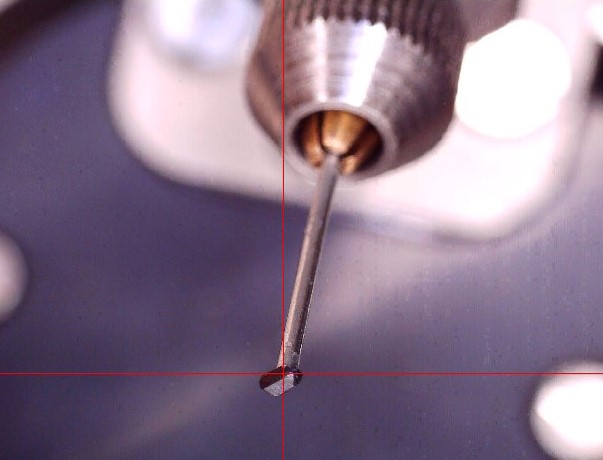
Objects of research
Documents
Video materials
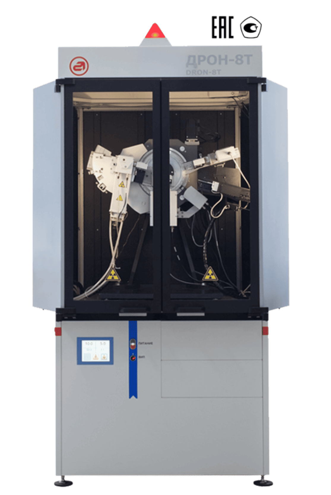
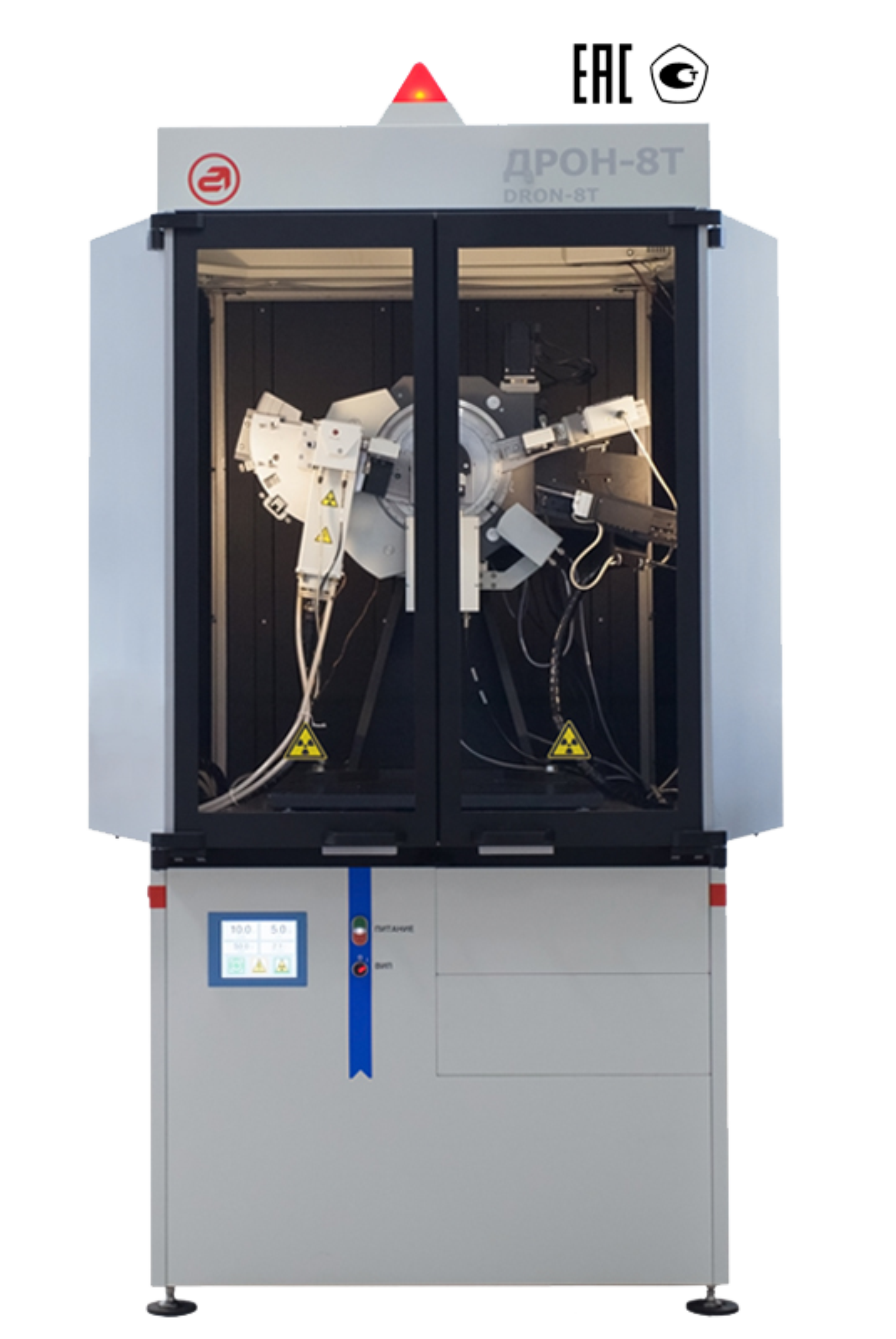
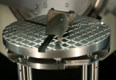
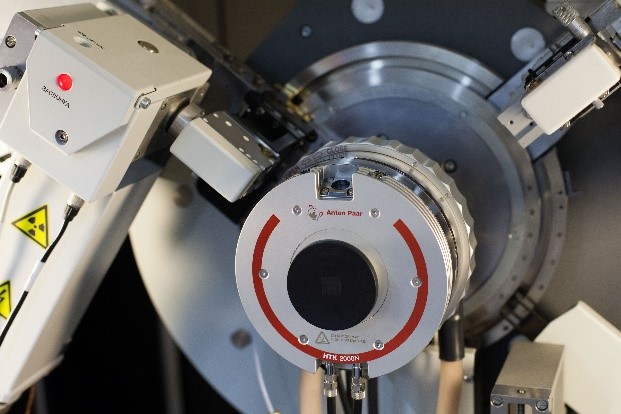
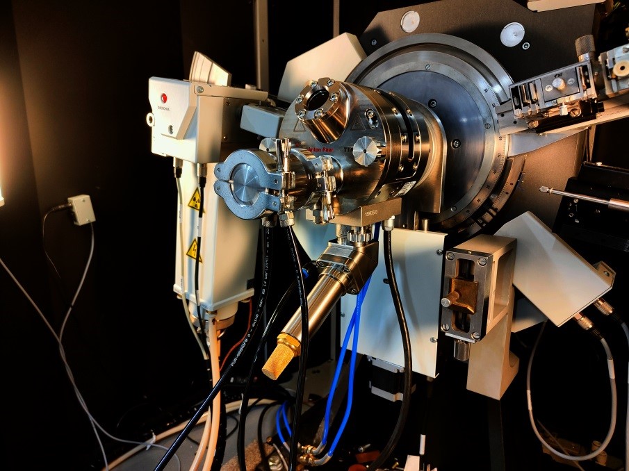
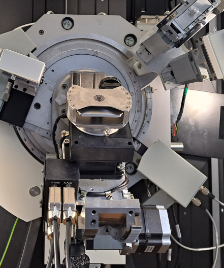
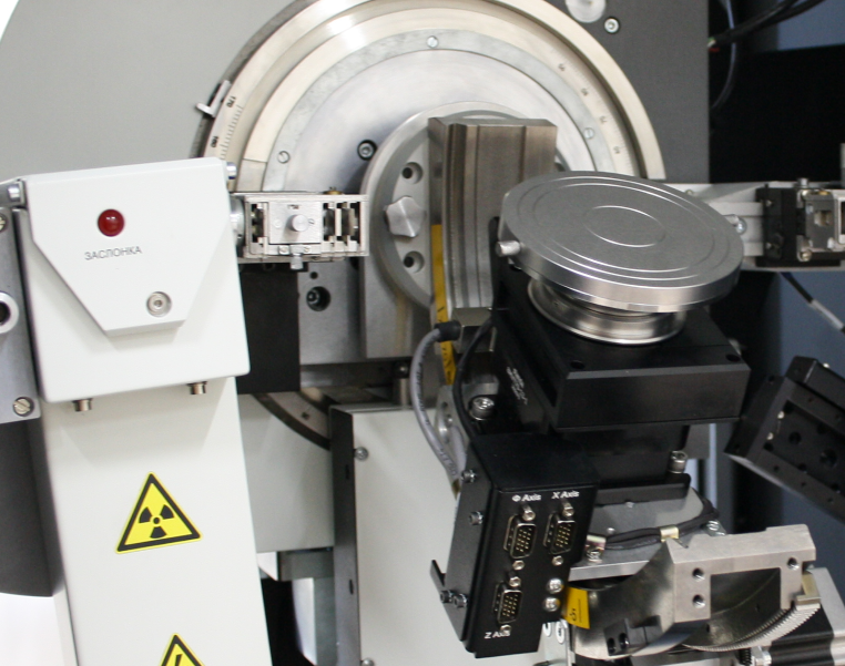
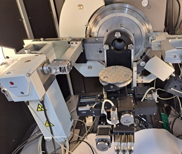
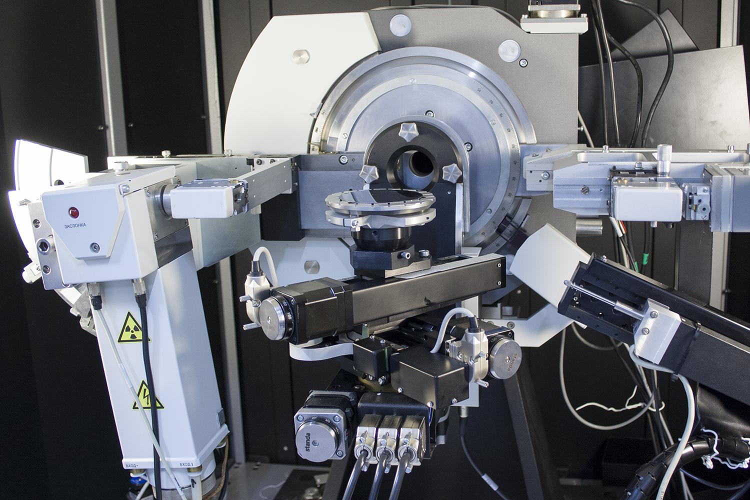
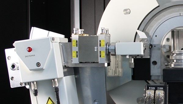
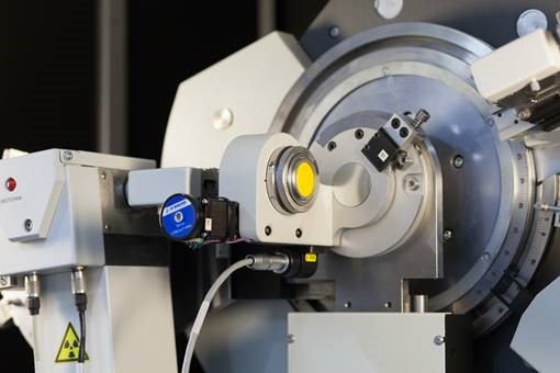
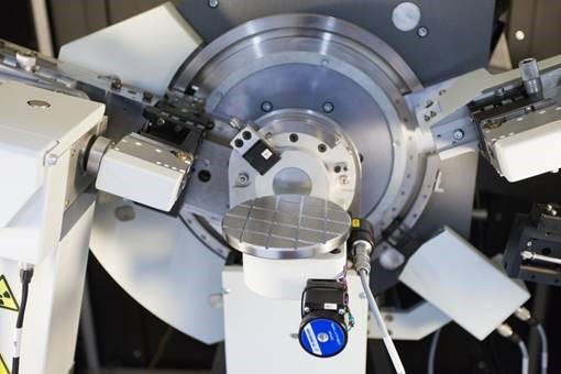
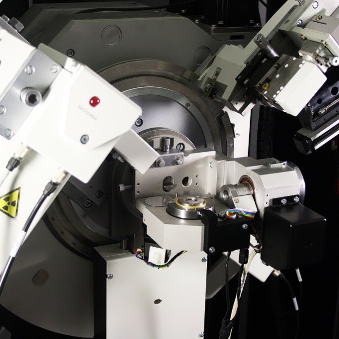
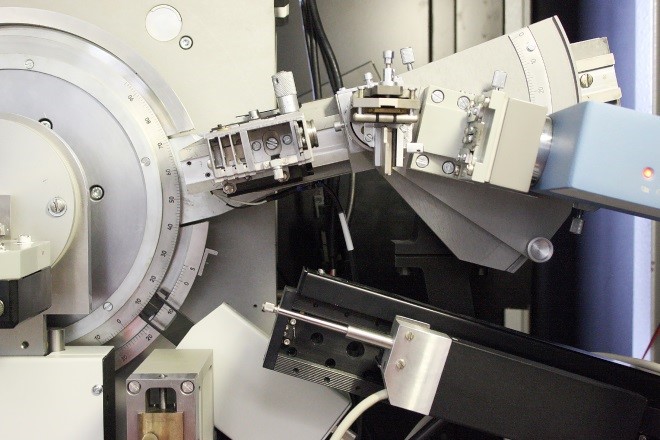
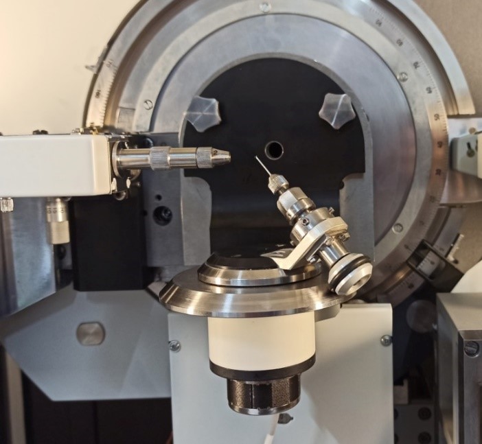
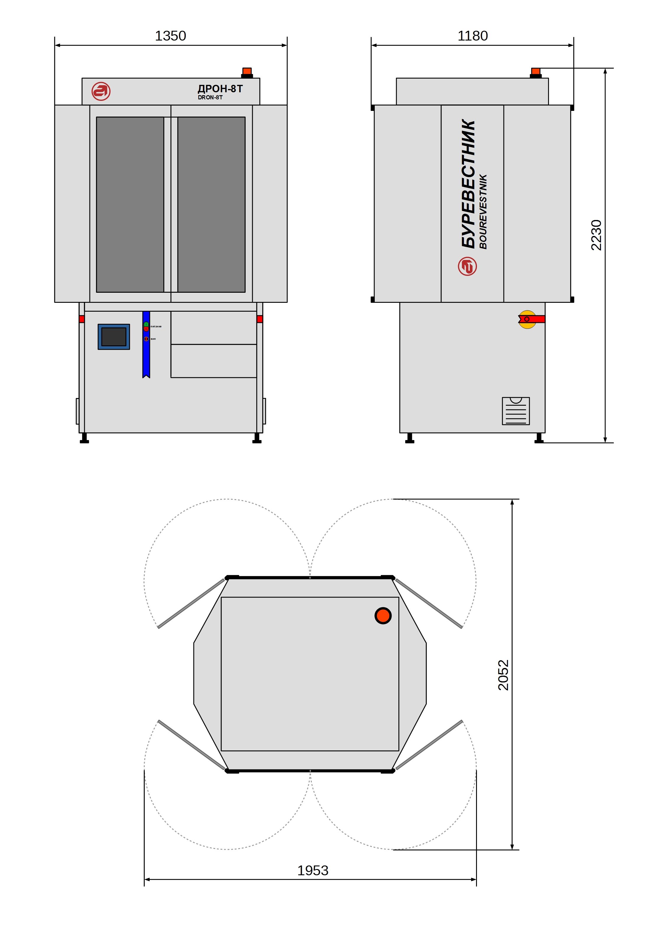
General-Purpose X-ray diffractometer DRON-8T
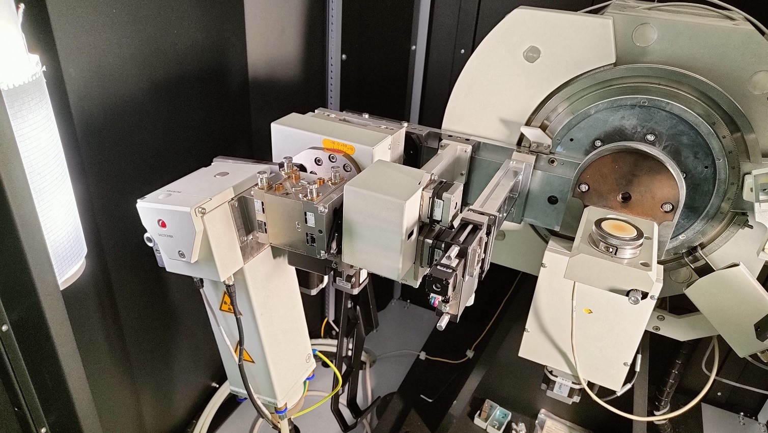
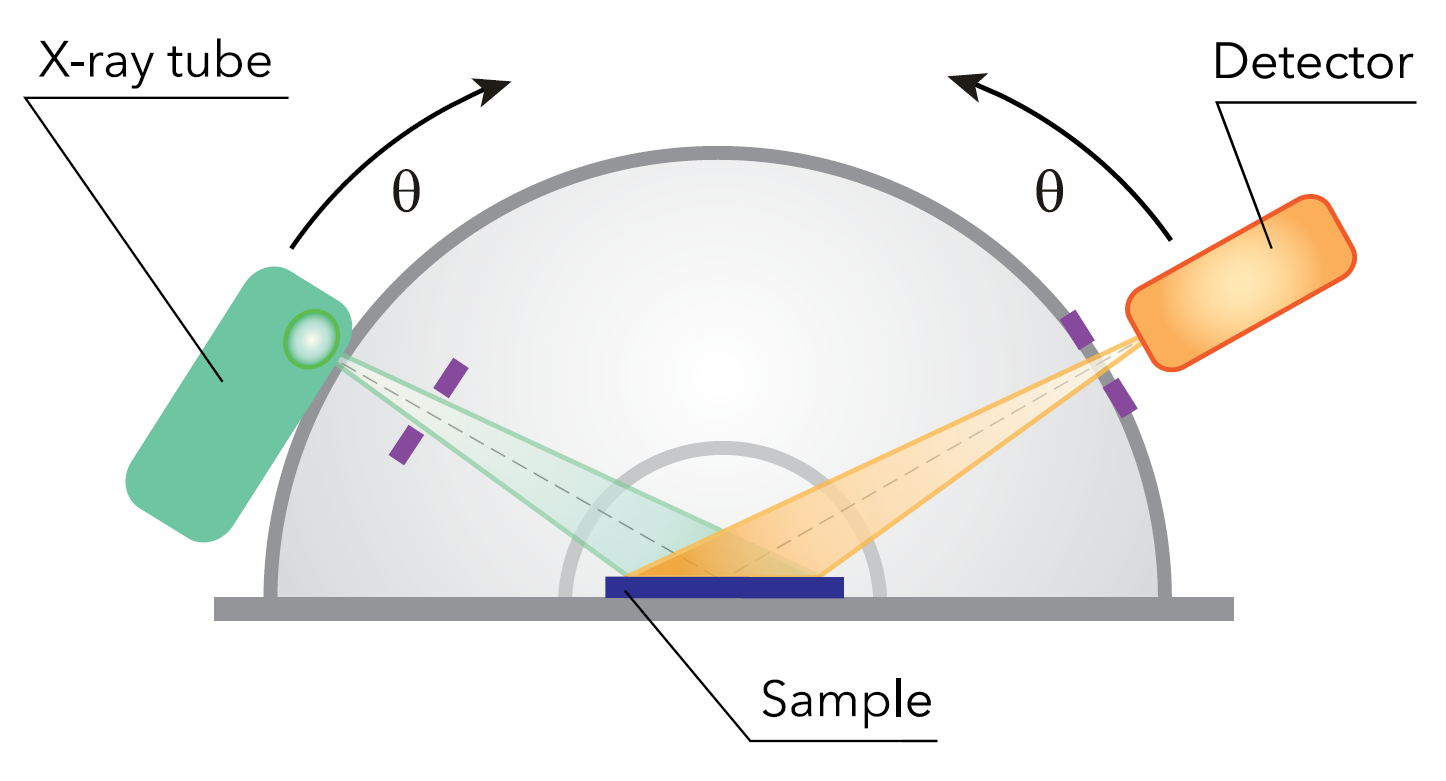
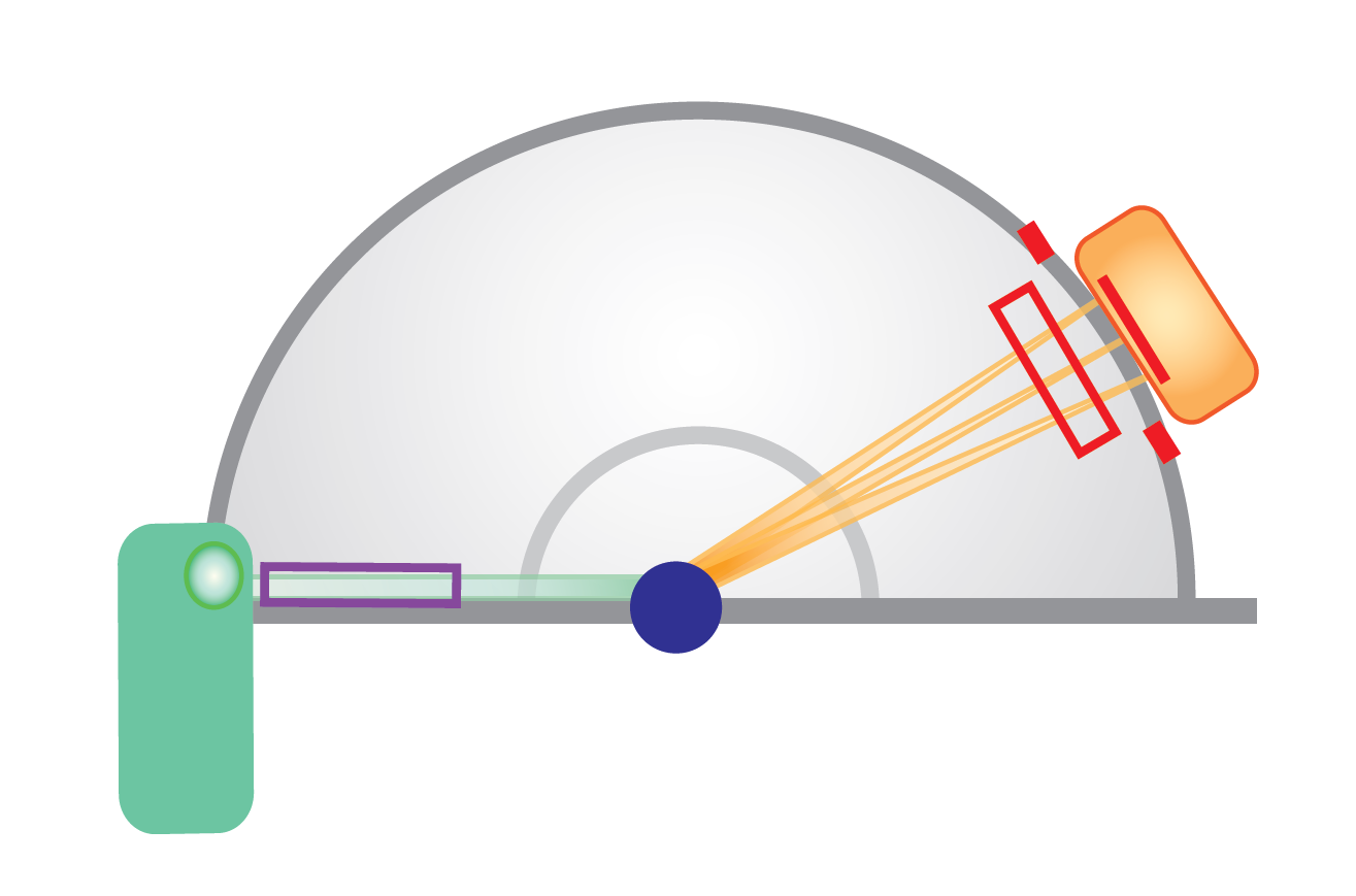
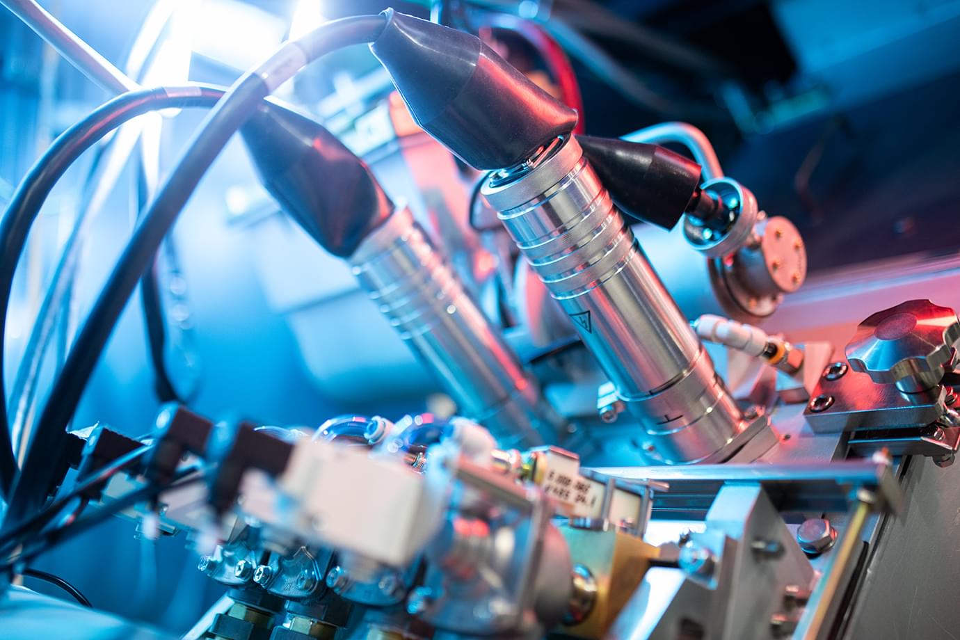
X-ray diffractometer DRON-8T is the flagship model of the line of stationary X-ray diffractometers of IC Bourevestnik JSC. It is equipped with a high-precision wide-angle vertical θ-θ goniometer with angular reproducibility of 0.0001° and is designed mainly for the study of highly refined crystalline objects (single crystals, epitaxial films) in high-resolution geometry.
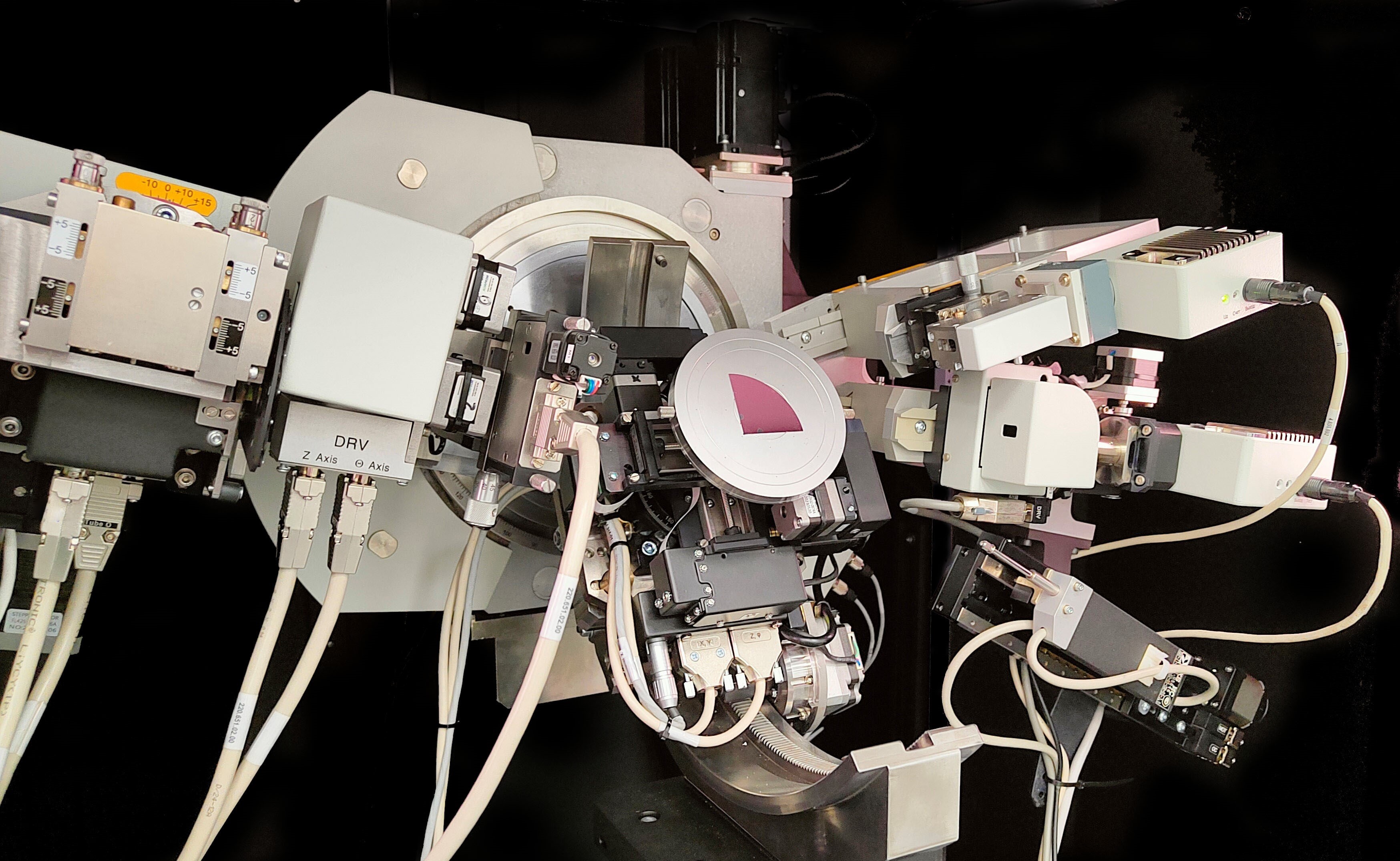
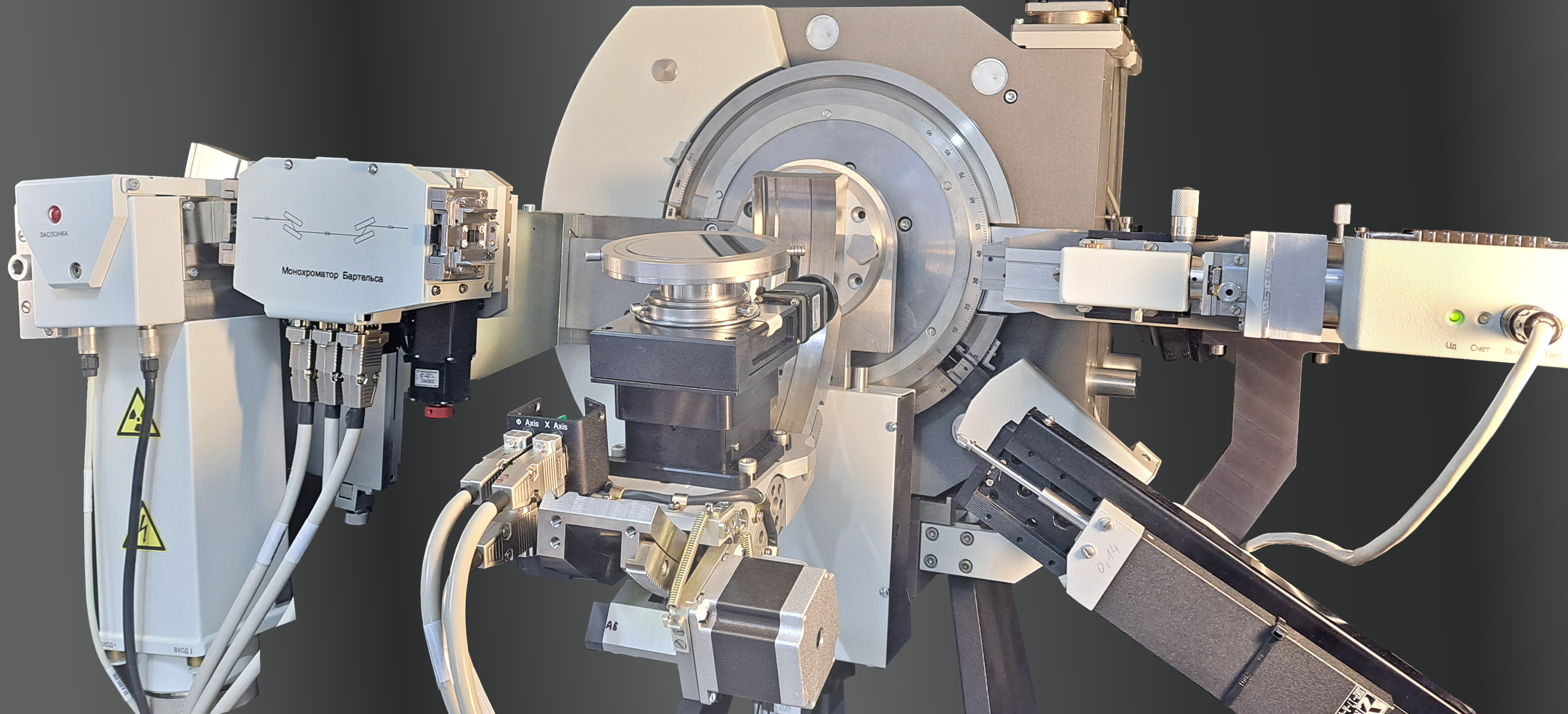

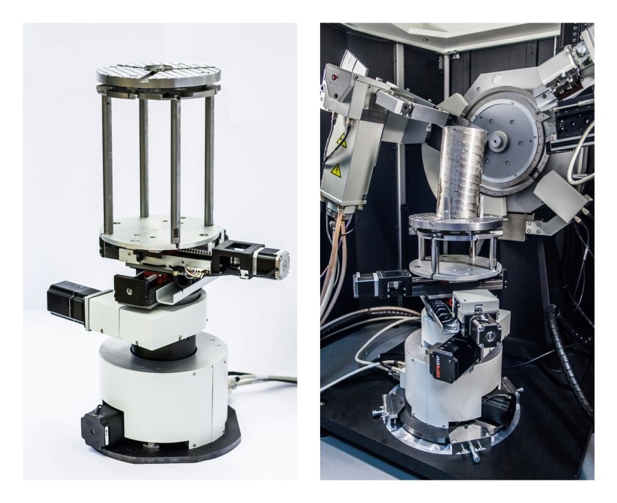
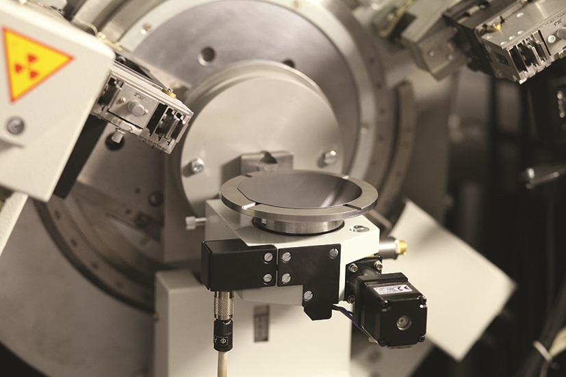
- High-precision wide-angle vertical goniometer with changeable radius
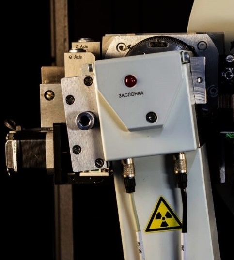
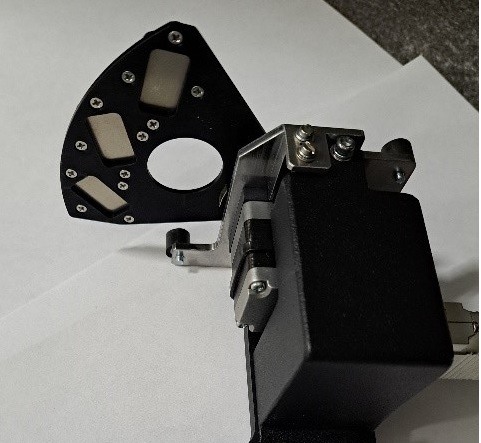
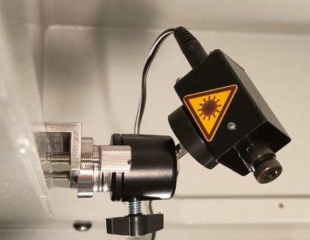
- Motorized tunable X-ray optical system (optional)
- Realization of different measurement methods
- Flexible machine configuration and a wide range of options
- Variety of X-ray optical schemes
- Software control of all devices and mechanisms


